Right Subacute Moderate S1 Radiculitis due to L5-S1 Central Protrusion, Complicated by Bilateral L4 Pars Defect (Clinical Case Review) (CR – 34)
Abstract
MRI studies were initially produced in the 1970s, and the first live man subject was imaged in 1977. Since that period, significant changes in both image quality and clinical performance have been achieved. Today, an MRI study can be obtained from any part of the body, with appreciable accuracy.
Its application in spinal bio-mechanics has also revolutionized our approach and treatment protocols for related disorders as well. More importantly, it has allowed visualization of specific morphology of the IVD structural pathology, an essential imaging advantage in performing closed decompression protocols.
Furthermore, being a relatively safe application, it has provided an excellent opportunity to monitor IVD structural pathology changes following treatment protocols.
Despite many advantages, MRI study limitations, particularly in respect to dynamic spinal bio-mechanics, should also be kept in mind.
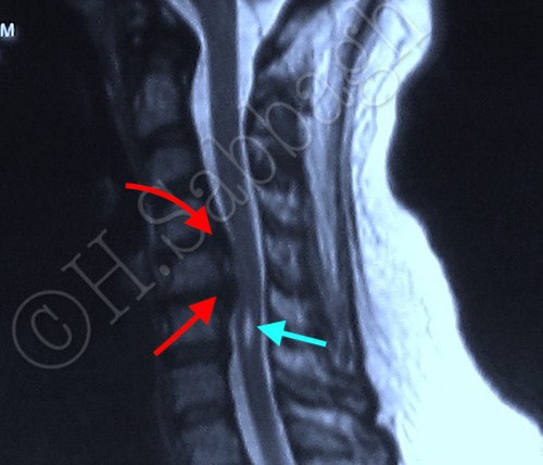
This clinical case review involves a patient who was diagnosed with a right subacute moderate S1 radiculitis, due to L5-S1 central protrusion.
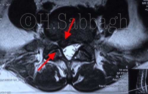
Even so, this patient underwent four separate MRI studies during a six-year period, yet the structural pars defect in the next cephalic segment was not reported, and the HSI in the pedicle was not visualized in any of the studies.
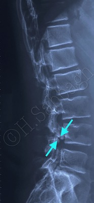
In addition, following the first three physical examinations, no request for complementary imaging studies such as CT or Stress studies was made, despite the patient’s clinical presentation suggesting chronic bio-mechanical instability.
In this CCR, serial MRI studies are presented, including the final study and the complimentary stress study results.
Further emphasis has been made on the importance of comprehensive physical examination, and bio-mechanical examination as an integrated component in spinal disorders. It is primarily the responsibility of the treating clinician to keep in mind that multiple bio-mechanical disorders can exist simultaneously, and the imaging study request must include the working diagnosis.
Spondylolysis is an important bio-mechanical finding which not only directly affected the treatment protocol of the L5-S1 IVD structural pathology, but it also alters the long term prognosis.